Upper Thigh Muscles Ct Anatomy : Описание: muscles_jambe5_ff_en | Piaaaach | Anatomia ... / Pain in the upper thigh can be difficult to diagnose because this area of the body contains many muscles, tendons, and ligaments.
Upper Thigh Muscles Ct Anatomy : Описание: muscles_jambe5_ff_en | Piaaaach | Anatomia ... / Pain in the upper thigh can be difficult to diagnose because this area of the body contains many muscles, tendons, and ligaments.. You've got an anterior compartment, medial, and posterior compartment and these are separated by the intermuscular this is this group of muscles here anteriorly in the thigh, obviously and these muscles are supplied by the femoral nerve. The thigh is the area between the hip and the knee joint. Muscle anatomy of upper thigh, human muscles, muscle anatomy of upper thigh. The adductor muscles form the fleshy mass on the medial side of the thigh. Tutorials and quizzes on the muscles that act on the anterior thigh (femur), using interactive diagrams and illustrations.
Learn faster with these free muscle labeling diagrams. The gluteus medius muscle helps abducts the thigh along with the gluteus maximus, but can rotate the thigh inward where the gluteus maximus rotates the write down the muscles of the thigh in the table below and, for each, give the location of that muscle and what effect contracting that muscle has. Pain in the upper thigh can be difficult to diagnose because this area of the body contains many muscles, tendons, and ligaments. The pectineus muscle is a flat muscle that forms the base of the femoral triangle. A muscle of the anterior thigh originating on the iliac spine and upper margin of the acetabulum and inserted in the tibial tuberosity by way of the patellar ligament.
Lower Limbs | Radiology Key from radiologykey.com This is my video about the muscles of the back. As the name implies they adduct the thigh at the hip joint. Learn faster with these free muscle labeling diagrams. In clinical anatomy the thigh muscles are divided into three groups: Written by keith bridwell, md; Thighs thigh muscles thigh actions and movements. A muscle of the medial thigh that originates on the pubis. It has a dual innervation, and thus can be considered a transitional.
Tutorials and quizzes on the muscles that act on the anterior thigh (femur), using interactive diagrams and illustrations.
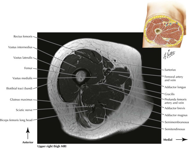
Pain in the upper thighlearn about different causes of upper thigh pain, from injuries to nerve problems. It inserts onto the linea aspera of the femur. This webpage presents the anatomical structures found on thigh mri. The iliopsoas is made up of two muscles that flex the thigh. We look at the associated symptoms and treatment options. Thigh muscle strains can occur when playing sports or participating in a daily activity. The single bone in the thigh is called the femur. In human anatomy, the thigh is the area between the hip (pelvis) and the knee. Muscles are named according to their shape, location, or a combination. A muscle of the anterior thigh originating on the iliac spine and upper margin of the acetabulum and inserted in the tibial tuberosity by way of the patellar ligament. Anatomical structures of the lower limb (hip, thigh, knee, leg, ankle and foot) and specific regions (compartment of the lower limb) are visible on cross section of the leg : Thigh muscle strains are common for people of all ages. Muscles that move the shoulder and arm include the trapezius and serratus anterior.
Muscle anatomy of upper thigh, human muscles, muscle anatomy of upper thigh. The hamstring muscles in the back of the thigh, the quadriceps muscles in the front, and the muscle strains usually happen when a muscle is stretched beyond its limit, tearing the muscle fibers. We look at the associated symptoms and treatment options. Muscles are groups of cells in the body that have the ability to contract and relax. Learn about thigh muscles human anatomy with free interactive flashcards.
Leg Muscle Diagram Anatomy Of The Thigh Very Best Example ... from i.pinimg.com The chewing muscles enable you to chew your food by moving the upper and lower teeth against one another. As the cursor is moved over a particular compartment of the lower. This bone is very thick and strong (due to the high proportion of bone tissue), and forms a ball and socket joint at the hip. This injury frequently occurs near the point where the. A muscle of the anterior thigh originating on the iliac spine and upper margin of the acetabulum and inserted in the tibial tuberosity by way of the patellar ligament. In the upper back region, the trapezius, rhomboid major, and levator scapulae muscles anchor the scapula and clavicle to the spines of several vertebrae and in addition to moving the arm and pectoral girdle, muscles of the chest and upper back work together as a group to support the vital process of. The muscles of the thigh are arranged into three compartments. Anatomically, it is part of the lower limb.
In human anatomy, the thigh is the area between the hip (pelvis) and the knee.
The muscle becomes stressed and tired after repeatedly doing the same motions over and over, leaving muscles fibers vulnerable to tears. This webpage presents the anatomical structures found on thigh mri. Superior ramus of the pubis insertion: The posterior compartment of the thigh contains the knee flexors and hip extensors.it has the following muscles, nerves and vessels: Tutorials and quizzes on the muscles that act on the anterior thigh (femur), using interactive diagrams and illustrations. Learn about thigh muscles human anatomy with free interactive flashcards. It is part of the lower limb. A muscle of the medial thigh that originates on the pubis. It crosses the back of the shoulder and attaches to the upper humeral shaft, below the head. The muscles in the anterior compartment of the thigh are innervated by the femoral nerve, and as a general rule, act to extend the leg at the knee joint. In human anatomy, the thigh is the area between the hip (pelvis) and the knee. This is my video about the muscles of the back. Thigh muscle strains can occur when playing sports or participating in a daily activity.
Pain in the upper thigh can be difficult to diagnose because this area of the body contains many muscles, tendons, and ligaments. Anatomically, it is part of the lower limb. The sparthos thigh compression sleeve provides compression as well as support for thigh muscles. Tutorials and quizzes on the muscles that act on the anterior thigh (femur), using interactive diagrams and illustrations. This injury frequently occurs near the point where the.
Figure 6 from Normal MR imaging anatomy of the thigh and ... from ai2-s2-public.s3.amazonaws.com Reviewed by mary rodts, dnp. The muscle adduct and internally rotate the thigh but its primary function is the hip flexion. 2, vastus medialis & intermedius muscles. The posterior compartment of the thigh contains the knee flexors and hip extensors.it has the following muscles, nerves and vessels: The muscle becomes stressed and tired after repeatedly doing the same motions over and over, leaving muscles fibers vulnerable to tears. Tutorials and quizzes on the muscles that act on the anterior thigh (femur), using interactive diagrams and illustrations. Muscles of the posterior cervical and upper thoracic spine 1. The thigh has three sets of strong muscles:
Muscle anatomy of upper thigh, human muscles, muscle anatomy of upper thigh.
Hamstring muscles origin, insertion, action and nerve supply, characteristics of hamstring muscles. The pectineus muscle is a flat muscle that forms the base of the femoral triangle. Anatomical structures of the lower limb (hip, thigh, knee, leg, ankle and foot) and specific regions (compartment of the lower limb) are visible on cross section of the leg : A muscle of the medial thigh that originates on the pubis. Muscles that move the shoulder and arm include the trapezius and serratus anterior. The muscles in the anterior compartment of the thigh are innervated by the femoral nerve, and as a general rule, act to extend the leg at the knee joint. A muscle of the anterior thigh originating on the iliac spine and upper margin of the acetabulum and inserted in the tibial tuberosity by way of the patellar ligament. Superior ramus of the pubis insertion: In human anatomy, the thigh is the area between the hip (pelvis) and the knee. It arises by tendinous fibers from the anterior superior iliac spine and the upper half of the notch below it. Muscles are named according to their shape, location, or a combination. Muscles of the posterior cervical and upper thoracic spine 1. The muscles of the thigh are arranged into three compartments.
Muscles are groups of cells in the body that have the ability to contract and relax upper thigh anatomy. As the cursor is moved over a particular compartment of the lower.